

Pathology and Tissue-Level Evaluation Support
Visualize what is happening in tissues when blood data or clinical observations alone cannot fully explain efficacy, safety, or mechanism of action.
KGAH-ONE works backward from the development question to organize the tissue, staining method, pathology endpoint, image quantification, and molecular or immune readouts, then connects the program with suitable specialized partners.
Design tissue evidence for a development decision—not just a set of representative images.
The meaning of the result can differ depending on which tissue region improved, how much injury, inflammation, fibrosis, or cellular infiltration changed, and whether those changes align with the intended mechanism.
KGAH-ONE organizes which tissues, stains, markers, scoring systems, and quantitative image methods should be used according to disease, mechanism, intervention timing, and the expected dog or cat study.
Can the observed efficacy, safety, or mechanism be explained at the tissue and cellular level?
Connect lesion location, severity, distribution, cell composition, fibrosis, and marker expression with the development question.
Combine morphology, pathology scoring, positive area, and marker localization to convert tissue change into decision-ready evidence.
The goal is not a single representative image, but an evaluation package that supports group comparison, candidate ranking, mechanism confirmation, and next-study design.
Four perspectives in pathology analysis
Go beyond whether a change exists, and define where it occurs, how extensive it is, and which tissue or cellular element is involved.
Tissue Structure and Injury
Evaluate pathology such as tubular injury, hepatocellular injury, alveolar injury, or tumor-tissue change.
Inflammation and Fibrosis
Combine inflammatory-cell infiltration, collagen deposition, fibrosis area, and markers such as α-SMA.
Cell and Marker Localization
Consider IHC / IF where appropriate to examine target, cell type, immune cells, or site of action.
Image Quantification and Group Comparison
Use positive area, fibrosis area, pathology score, or stage to compare candidates, doses, and groups.
Representative pathology images approved for public use
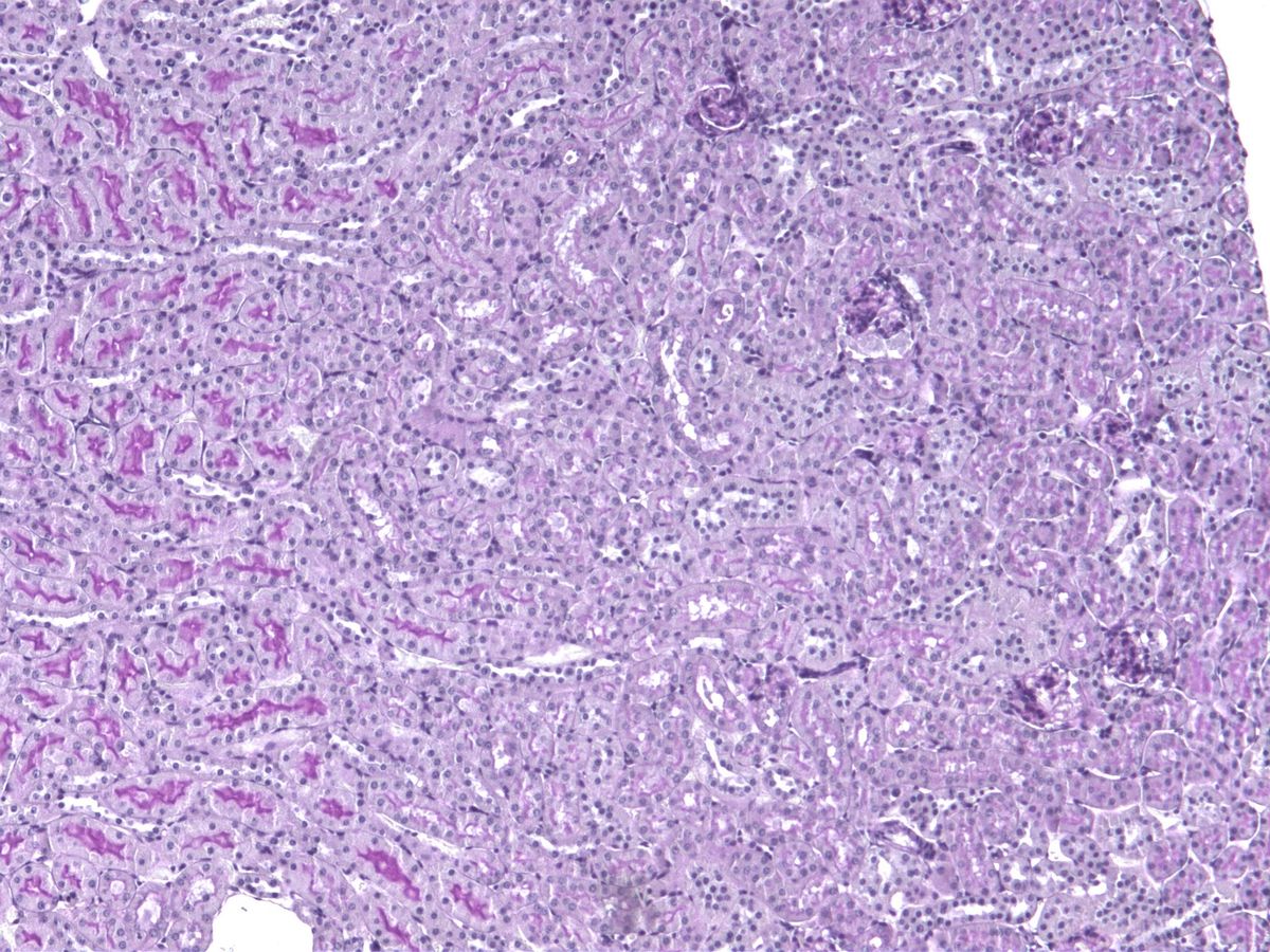
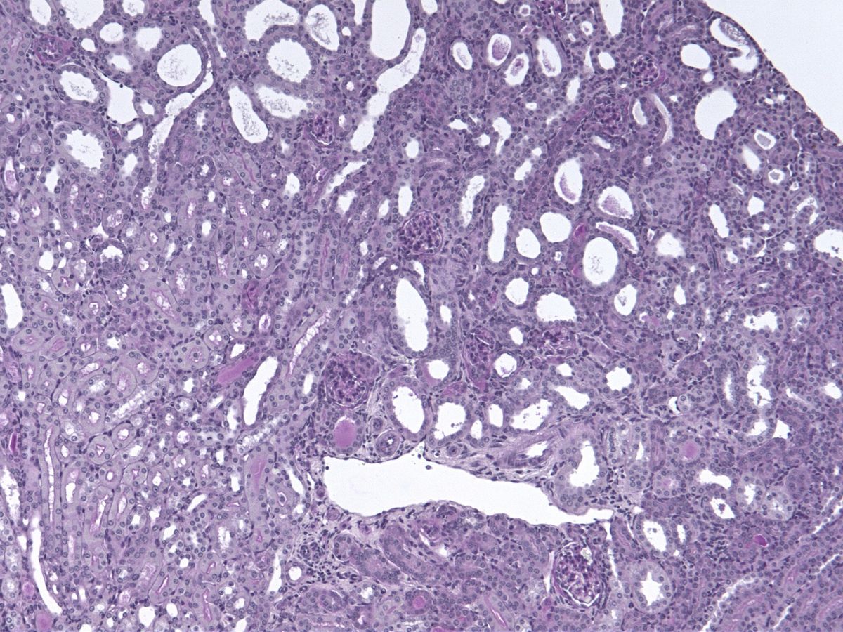
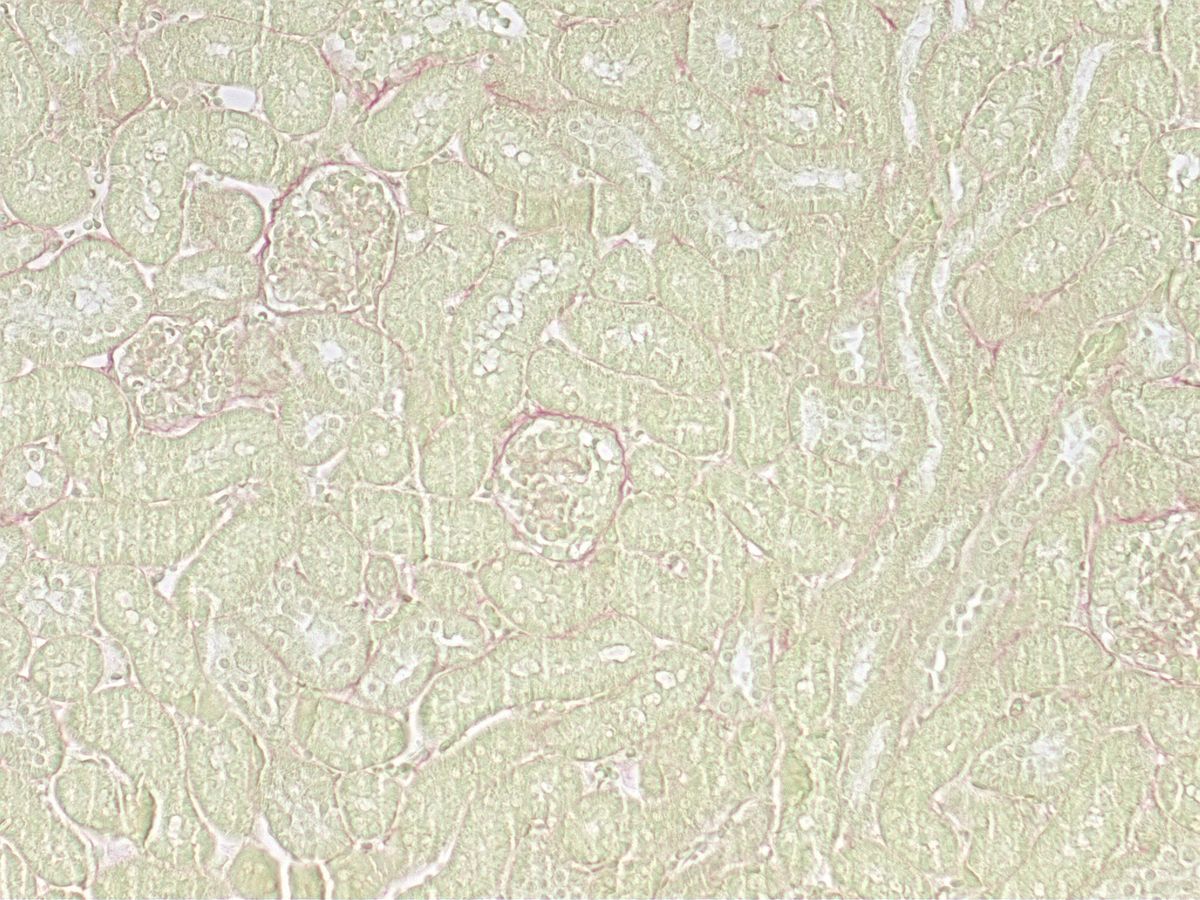
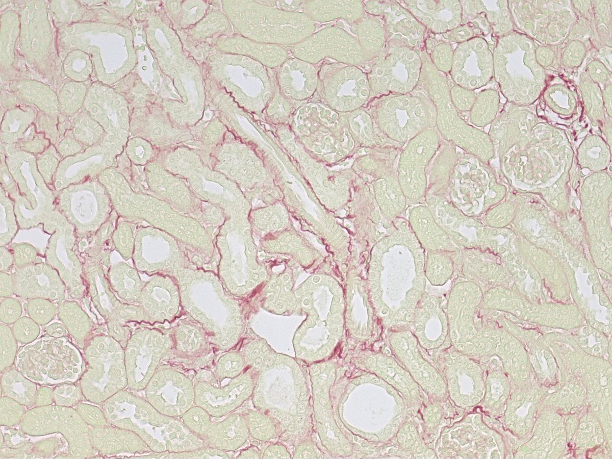
These representative images are from an adenine-induced kidney injury model. PAS staining visualizes tubulointerstitial injury, while Sirius Red staining highlights renal fibrosis and collagen deposition.
PAS Staining — Tubulointerstitial Injury
Representative comparison of normal kidney tissue and adenine-induced kidney injury
Sirius Red Staining — Renal Fibrosis
Representative comparison of collagen deposition in normal kidney and adenine-induced kidney injury
These are representative images approved for public use. The stain, marker, observation area, magnification, and quantification method should be designed according to the disease, mechanism, sample, and development question.
Convert pathology images into quantitative evidence
Avoid drawing conclusions from representative images alone. Combine pathology with disease-relevant scoring and quantitative readouts.
Development Question
Define what pathology must answer: efficacy, safety, mechanism, or candidate comparison.
Tissue, Stain, and Marker
Select the target organ, lesion area, PAS, Sirius Red, H&E, and IHC / IF options as appropriate.
Observation, Score, and Quantification
Evaluate tubulointerstitial injury, positive area, fibrosis stage, cellular infiltration, or related endpoints.
Development Decision
Connect the results with candidate ranking, dose, additional validation, Go / No-Go, or next dog and cat study design.
Representative application areas and pathology readouts
Combine tissue morphology, staining, image quantification, and molecular or immune markers according to the development objective.
Renal Disease
PAS-based tubulointerstitial injury, Sirius Red-positive area, α-SMA, collagen, and macrophage infiltration.
Liver Disease and MASH
Steatosis, lobular inflammation, ballooning, NAS, fibrosis stage, Sirius Red, and α-SMA.
Pulmonary Fibrosis and ILD
Ashcroft score, Sirius Red / Masson, α-SMA, and histological endpoints related to inflammation and fibrosis.
Oncology and Immuno-Oncology
H&E, IHC, proliferation / apoptosis markers, CD8 / macrophage infiltration, and PD-L1.
Representative public track record of a specialized partner
These are externally usable performance statements from SMC Laboratories. KGAH-ONE leads problem definition and coordination design.
Turn tissue-level change into the next development decision.
Tell us the target disease, sample, mechanism, current endpoints, and the tissue change or marker you need to clarify. KGAH-ONE can help organize pathology endpoints, staining, image quantification, molecular and immune readouts, and the specialized partner pathway.
KGAH-ONE is an Animal Health intelligence and partner coordination platform of Kawasaki Global Animal Health, Inc. Specialized pathology, nonclinical evaluation and scientific support may be provided by partners such as SMC Laboratories, depending on the development question.